

BRAKE & CONSIDER

MET overexpression and amplification are the most common drivers of progression after a 1L third-generation EGFR TKI1-3
ASSESS & DETECT

Tissue rebiopsy is critical in identifying MET alterations and is more sensitive than plasma alone2,3
GO FORWARD

Future novel treatment approaches may be able to address acquired MET drivers in patients with EGFRm NSCLC4-7
Understand what’s driving their progression. Tissue rebiopsy at the first sign of progression.
What’s driving progression?
Which mutations or changes lead to progression?
MET overexpression and amplification are the most common drivers of progression on third-generation EGFR TKIs1-3
Histologic transformation (eg, SCLC) is the second most common acquired driver1-3*
Patients treated with a third-generation EGFR TKI
generally will acquire new drivers of progression2
MET overexpression or amplification4

 of patients
- Up to ~34% of patients can have high levels of MET overexpression and/or amplification (IHC90+ and/or FISH10+)4
- Acquired abnormal activation of the MET pathway—including protein overexpression and gene amplification—is believed to be a dysregulator in carcinogenesis and is associated with a poor prognosis3,8
Histologic transformation (eg, SCLC)2

 of patients
- Histological changes—such as the transformation from NSCLC to SCLC—require a different treatment approach9
NCCN Clinical Practice Guidelines in Oncology (NCCN Guidelines®) recommend considering a tissue rebiopsy at time of progression to rule out SCLC transformation and understand drivers of progression.10
*Other acquired drivers of progression include: KRAS amplification/mutation (3%-4%), PIK3CA (2%-3%), BRAF V600E (2%), ALK fusion (1%-2%), BRAF fusion (≤1%), HER2 amplification (≤1%), HER3 amplification (≤1%), and RET fusion (≤1%).2
Why tissue biopsy?
How to identify MET overexpression and amplification
Tissue rebiopsy is critical in identifying acquired MET-driven progression and is more sensitive than plasma alone2
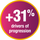
Drivers of progression, like MET amplification, were detected in 31% more cases with tissue biopsy than liquid biopsy2
- These results are from an analysis of 193 patients with advanced EGFRm NSCLC who had progressed on a third-generation EGFR TKI, including 58 patients who had both tissue and liquid biopsies*

MET overexpression is only detected by tissue biopsy/IHC3
- Liquid biopsy relies on ctDNA and is not suitable for detection of protein-based biomarkers such as MET overexpression
In the TATTON study, NGS failed to detect approximately 50% of patients with MET overexpression or amplification11
- IHC and FISH on tissue rebiopsy are the most effective techniques to identify acquired MET drivers after 1L progression
Capture the full picture. Tissue rebiopsy at the first sign of progression.
ctDNA=circulating tumour DNA; FISH=fluorescence in situ hybridisation; IHC=immunohistochemistry; NGS=next-generation sequencing.
*Molecular testing was performed via NGS and FISH.2
Ongoing investigations
Ongoing clinical investigations
Combining MET and EGFR TKIs may be beneficial in treating EGFRm NSCLC after first-line progression
A number of ongoing clinical studies are investigating MET and EGFR TKI combinations in patients with advanced EGFRm NSCLC who have acquired MET drivers of progression following first-line treatment with a third-generation EGFR TKI4-7
Visit astrazeneca.com for more information.
1L=first-line; ALK=anaplastic lymphoma kinase; BRAF=v-raf murine sarcoma viral oncogene homolog B1; ctDNA=circulating tumour DNA; EGFR=epidermal growth factor receptor; EGFRm=epidermal growth factor receptor mutation; FISH=fluorescence in situ hybridisation; HER2=human epidermal growth factor receptor 2; HER3=human epidermal growth factor receptor 3; IHC=immunohistochemistry; KRAS=V-Ki-ras2 Kirsten rat sarcoma 2 viral oncogene homolog; MET=mesenchymal-epithelial transition; mNSCLC=metastatic non-small cell lung cancer; NCCN=National Comprehensive Cancer Network® (NCCN®); NGS=next-generation sequencing; NSCLC=non-small cell lung cancer; PIK3CA=phosphatidylinositol-4,5-bisphosphate 3-kinase catalytic subunit alpha; RET=rearranged during transfection; SCLC=small cell lung cancer; TKI=tyrosine kinase inhibitor.
References: 1. Choudhury NJ, Marra A, Sui JSY, et al. Molecular biomarkers of disease outcomes and mechanisms of acquired resistance to first-line osimertinib in advanced EGFR-mutant lung cancers. J Thorac Oncol. 2022;18(4):463-475. 2. Piotrowska Z, Chen LN, Shum E, et al. Tissue and plasma-based mechanisms of resistance to first-line osimertinib in EGFR-mutant NSCLC: a multi-institutional cohort. Poster presented at: American Society of Clinical Oncology Annual Meeting; June 2-6, 2023; Chicago, IL. Poster 9108. 3. Bai Q, Shi X, Zhou X, et al. Chinese expert consensus on clinical practice of MET detection in non-small cell lung cancer. Ther Adv Med Oncol. 2024;16:1-19. 4. Ahn MJ, DeMarinis F, Bonanno L, et al. MET biomarker-based preliminary efficacy analysis in SAVANNAH: savolitinib+osimertinib in EGFRm NSCLC post-osimertinib. Poster presented at: World Conference on Lung Cancer; August 6-9, 2022; Vienna, Austria. Poster #EP08.02-140. 5. Study on savolitinib combined with osimertinib in treatment of advanced NSCLC with MET amplification (SACHI). ClinicalTrials.gov identifier: NSCT05015608. Updated March 30, 2023. Accessed January 24, 2025. https://clinicaltrials.gov/study/NCT05015608. 6. Savolitinib plus osimertinib versus platinum-based doublet chemotherapy in participants with non-small cell lung cancer who have progressed on osimertinib treatment (SAFFRON). ClinicalTrials.gov identifier: NCT05261399. Updated January 10, 2025. Accessed January 24, 2025. https://clinicaltrials.gov/study/NCT05261399. 7. Phase 2 platform study in patients with advanced non-small lung cancer who progressed on first-line osimertinib therapy (ORCHARD). ClinicalTrials.gov identifier: NCT03944772. Updated January 30, 2025. Accessed January 30, 2025. https://www.clinicaltrials.gov/study/NCT03944772. 8. Tsuta K, Kozu Y, Mimae T, et al. c-MET/phospho-MET protein expression and MET gene copy number in non-small cell lung carcinomas. J Thorac Oncol. 2012;7(2):331-339. 9. Yang Y, Fan S. Small cell lung cancer transformations from non-small cell lung cancer: biological mechanism and clinical relevance. Chin Med J Pulm Crit Care Med. 2024;2(1):42-47. 10. Referenced with permission from the NCCN Clinical Practice Guidelines in Oncology (NCCN Guidelines®) for Non-Small Cell Lung Cancer V.4.2025. © National Comprehensive Cancer Network, Inc. 2025. All rights reserved. Accessed May 23, 2025. To view the most recent and complete version of the guideline, go online to NCCN.org. NCCN makes no warranties of any kind whatsoever regarding their content, use or application and disclaims any responsibility for their application or use in any way. 11. Hartmaier RJ, Markovets AA, Ahn MJ, et al. Osimertinib + savolitinib to overcome acquired MET-mediated resistance in epidermal growth factor receptor-mutated, MET-amplified non-small cell lung cancer: TATTON. Cancer Discov. 2023;13(1):98-113.
This site is intended for healthcare professionals only.
This site is fully developed and sponsored by AstraZeneca.
©2025 AstraZeneca. All rights reserved. Z4-74271 June 2025
